Προβολές σελίδας

Παρασκευή 21 Νοεμβρίου 2014

                                            Smallpox

                                  File:Smallpox virus virions TEM PHIL 1849.JPG
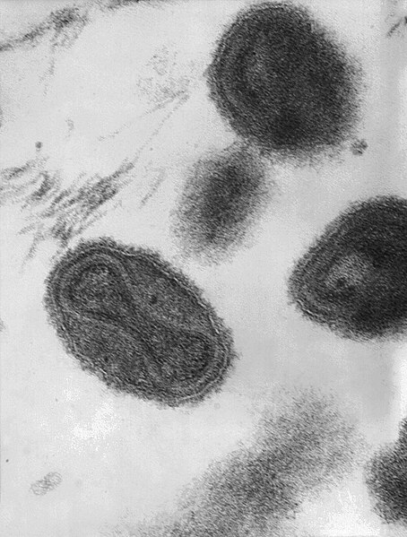
Smallpox was an infectious disease caused by either of two virus variants, Variola major and Variola minor.
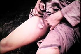
Smallpox was localized in small blood vessels of the skin and in the mouth and throat. In the skin it resulted in a characteristic maculopapular rash and, later, raised fluid-filled blisters. V. major produced a more serious disease and had an overall mortality rate of 30–35%. V. minor caused a milder form of disease, which killed about 1% of its victims. Long-term complications of V. major infection included characteristic scars, commonly on the face, which occur in 65–85% of survivors. Blindness resulting from corneal ulceration and scarring, and limb deformities due to arthritis and osteomyelitis were less common complications, seen in about 2–5% of cases.Smallpox is believed to have emerged in human populations about 10,000 BC. The earliest physical evidence of it is probably the pustular rash on the mummified body of Pharaoh Ramses V of Egypt. The disease killed an estimated 400,000 Europeans annually during the closing years of the 18th century (including five reigning monarchs), and was responsible for a third of all blindness. Of all those infected, 20–60%—and over 80% of infected children—died from the disease. Smallpox was responsible for an estimated 300–500 million deaths during the 20th century. As recently as 1967, the World Health Organization (WHO) estimated that 15 million people contracted the disease and that two million died in that year.


                                                       Signs and symptoms:
The incubation period between contraction and the first obvious symptoms of the disease is around 12 days. Once inhaled, variola major virus invades the oropharyngeal (mouth and throat) or the respiratory mucosa, migrates to regional lymph nodes, and begins to multiply. In the initial growth phase the virus seems to move from cell to cell, but around the 12th day, lysis of many infected cells occurs and the virus is found in the bloodstream in large numbers and a second wave of multiplication occurs in the spleen, bone marrow, and lymph nodes. The initial or prodromal symptoms are similar to other viral diseases such as influenza and the common cold: fever of at least 38.5 °C (101 °F), muscle pain, malaise, headache and prostration. As the digestive tract is commonly involved, nausea and vomiting and backache often occur. The prodrome, or preeruptive stage, usually lasts 2–4 days. By days 12–15 the first visible lesions—small reddish spots called enanthem—appear on mucous membranes of the mouth, tongue, palate, and throat, and temperature falls to near normal. These lesions rapidly enlarge and rupture, releasing large amounts of virus into the saliva
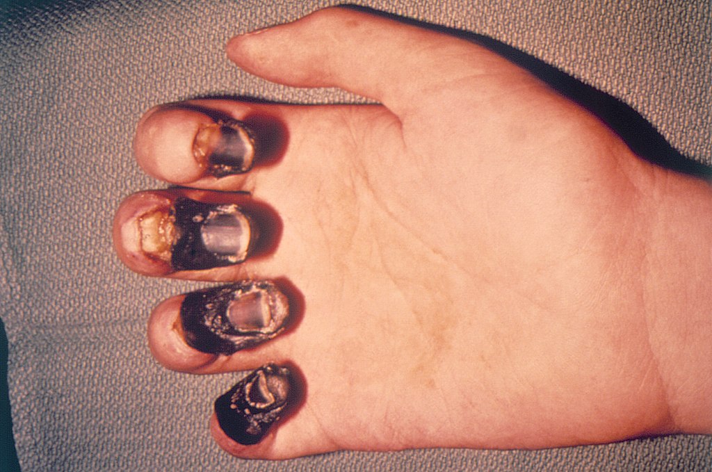


                                                                Hemorrhagic:

Hemorrhagic smallpox is a severe form that is accompanied by extensive bleeding into the skin, mucous membranes, and gastrointestinal tract. This form develops in approximately 2% of infections and occurred mostly in adults. In hemorrhagic smallpox the skin does not blister, but remains smooth. Instead, bleeding occurs under the skin, making it look charred and black, hence this form of the disease is also known as black pox.


                                                         Transimission:
Transmission occurs through inhalation of airborne variola virus, usually droplets expressed from the oral, nasal, or pharyngeal mucosa of an infected person. It is transmitted from one person to another primarily through prolonged face-to-face contact with an infected person, usually within a distance of 6 feet (1.8 m), but can also be spread through direct contact with infected bodily fluids or contaminated objects (fomites) such as bedding or clothing. Rarely, smallpox has been spread by virus carried in the air in enclosed settings such as buildings, buses, and trains. Smallpox is highly contagious, but generally spreads more slowly and less widely than some other viral diseases, perhaps because transmission requires close contact and occurs after the onset of the rash.

                                                         Treatment:
Smallpox vaccination within three days of exposure will prevent or significantly lessen the severity of smallpox symptoms in the vast majority of people. Vaccination four to seven days after exposure can offer some protection from disease or may modify the severity of disease. Other than vaccination, treatment of smallpox is primarily supportive, such as wound care and infection control, fluid therapy, and possible ventilator assistance. Flat and hemorrhagic types of smallpox are treated with the same therapies used to treat shock, such as fluid resuscitation. People with semi-confluent and confluent types of smallpox may have therapeutic issues similar to patients with extensive skin burns.No drug is currently approved for the treatment of smallpox. However, antiviral treatments have improved since the last large smallpox epidemics, and studies suggest that the antiviral drug cidofovir might be useful as a therapeutic agent. The drug must be administered intravenously, however, and may cause serious kidney toxicity.


Παρασκευή 7 Νοεμβρίου 2014

                                                         The Black Death:

The Black Death was one of the most devastating pandemics in human history, resulting in the deaths of an estimated 75 to 200 million people and peaking in Europe in the years 1346–53. Although there were several competing theories as to the etiology of the Black Death, analysis of DNA from victims in northern and southern Europe published in 2010 and 2011 indicates that the pathogen responsible was the Yersinia pestis bacterium, probably causing several forms of plague.

                                                     Symptoms:

Contemporary accounts of the plague are often varied or imprecise. The most commonly noted symptom was the appearance of buboes (or gavocciolos) in the groin, the neck and armpits, which oozed pus and bled when opened.
This was followed by acute fever and vomiting of blood. Most victims died two to seven days after initial infection. David Herlihy identifies freckle-like spots and rashes which could be caused by flea-bites as another potential sign of the plague.

A hand showing how arcal gangrene of the fingers due to bubonic plague causes the skin and flesh to die and turn black.


                                                       Causes:


The dominant explanation for the Black Death is the plague theory, which attributes the outbreak to Yersinia pestis, also responsible for an epidemic that began in southern China in 1865, eventually spreading to India. The investigation of the pathogen that caused the 19th-century plague was begun by teams of scientists who visited Hong Kong in 1894, among whom was the French-Swiss bacteriologist Alexandre Yersin, after whom the pathogen was named Yersinia pestis. The mechanism by which Y. pestis was usually transmitted was established in 1898 by Paul-Louis Simond and was found to involve the bites of fleas whose midguts had become obstructed by replicating Y. pestis several days after feeding on an infected host. This blockage results in starvation and aggressive feeding behaviour by the fleas, which repeatedly attempt to clear their blockage by regurgitation, resulting in thousands of plague bacteria being flushed into the feeding site, infecting the host. The bubonic plague mechanism was also dependent on two populations of rodents: one resistant to the disease, which act as hosts, keeping the disease endemic; and a second that lack resistance. When the second population dies, the fleas move on to other hosts, including people, thus creating a human epidemic.


                   The overall spread of the Black Death in Europe


Παρασκευή 31 Οκτωβρίου 2014

                                       Prions:

File:Histology bse.jpg


Prions are responsible for the transmissible spongiform encephalopathies in a variety of mammals, including bovine spongiform encephalopathy (BSE, also known as "mad cow disease") in cattle. In humans, prions cause Creutzfeldt-Jakob Disease (CJD), variant Creutzfeldt-Jakob Disease (vCJD), Gerstmann–Sträussler–Scheinker syndrome, Fatal Familial Insomnia and kuru. All known prion diseases in mammals affect the structure of the brain or other neural tissue and all are currently untreatable and universally fatal.
Prions are not considered living organisms but are misfolded protein molecules which may propagate by transmitting a misfolded protein state. If a prion enters a healthy organism, it induces existing, properly folded proteins to convert into the disease-associated, misfolded prion form; the prion acts as a template to guide the misfolding of more proteins into prion form. These newly formed prions can then go on to convert more proteins themselves; this triggers a chain reaction that produces large amounts of the prion form. All known prions induce the formation of an amyloid fold, in which the protein polymerises into an aggregate consisting of tightly packed beta sheets. Amyloid aggregates are fibrils, growing at their ends, and replicating when breakage causes two growing ends to become four growing ends. The incubation period of prion diseases is determined by the exponential growth rate associated with prion replication, which is a balance between the linear growth and the breakage of aggregates.
Prions cause neurodegenerative disease by aggregating extracellularly within the central nervous system to form plaques known as amyloid, which disrupt the normal tissue structure. This disruption is characterized by "holes" in the tissue with resultant spongy architecture due to the vacuole formation in the neurons. Other histological changes include astrogliosis and the absence of an inflammatory reaction. While the incubation period for prion diseases is relatively long (5 to 20 years), once symptoms appear the disease progresses rapidly, leading to brain damage and death.Neurodegenerative symptoms can include convulsions, dementia, ataxia (balance and coordination dysfunction), and behavioural or personality changes.


                                                         Transmission:
It has been recognized that prion diseases can arise in three different ways: acquired, familial, or sporadic. It is often assumed that the diseased form directly interacts with the normal form to make it rearrange its structure. One idea, the "Protein X" hypothesis, is that an as-yet unidentified cellular protein (Protein X) enables the conversion of PrPC to PrPSc by bringing a molecule of each of the two together into a complex.
Current research suggests that the primary method of infection in animals is through ingestion. It is thought that prions may be deposited in the environment through the remains of dead animals and via urine, saliva, and other body fluids. They may then linger in the soil by binding to clay and other minerals.

                                                           Sterilization:
Infectious particles possessing nucleic acid are dependent upon it to direct their continued replication. Prions, however, are infectious by their effect on normal versions of the protein. Sterilizing prions, therefore, requires the denaturation of the protein to a state in which the molecule is no longer able to induce the abnormal folding of normal proteins. In general, prions are quite resistant to proteases, heat, radiation, and formalin treatments, although their infectivity can be reduced by such treatments. Effective prion decontamination relies upon protein hydrolysis or reduction or destruction of protein tertiary structure. Examples include bleach, caustic soda, and strongly acidic detergents such as LpH. 134 °C (274 °F) for 18 minutes in a pressurized steam autoclave may not be enough to deactivate the agent of disease. Ozone sterilization is currently being studied as a potential method for prion denaturation and deactivation. Renaturation of a completely denatured prion to infectious status has not yet been achieved; however, partially denatured prions can be renatured to an infective status under certain artificial conditions
The World Health Organization recommends any of the following three procedures for the sterilization of all heat-resistant surgical instruments to ensure that they are not contaminated with prions:

Immerse in a pan containing 1N NaOH and heat in a gravity-displacement autoclave at 121 °C for 30 minutes; clean; rinse in water; and then perform routine sterilization processes.
Immerse in 1N NaClO (sodium hypochlorite) (20,000 parts per million available chlorine) for 1 hour; transfer instruments to water; heat in a gravity-displacement autoclave at 121 °C for 1 hour; clean; and then perform routine sterilization processes.
Immerse in 1N NaOH or sodium hypochlorite (20,000 parts per million available chlorine) for 1 hour; remove and rinse in water, then transfer to an open pan and heat in a gravity-displacement (121 °C) or in a porous-load (134 °C) autoclave for 1 hour; clean; and then perform routine sterilization processes.


                                             Parasites:

Parasitism is a non-mutual symbiotic relationship between species, where one species, the parasite, benefits at the expense of the other, the host. Traditionally parasite referred primarily to organisms visible to the naked eye, or macroparasites.Parasite now includes microparasites, which are typically smaller, such as protozoans, viruses and bacteria.Unlike predators, parasites do not kill their host, are generally much smaller than their host, and will often live in or on their host for an extended period. Both are special cases of consumer-resource interactions. Parasites show a high degree of specialization, and reproduce at a faster rate than their hosts. Classic examples of parasitism include interactions between vertebrate hosts and tapeworms, flukes, the Plasmodium species, and fleas. Parasitism differs from the parasitoid relationship because parasitoids generally kill their hosts.(A parasitoid is an organism that spends a significant portion of its life history attached to or within a single host organism in a relationship that is in essence parasitic; unlike a true parasite, however, it ultimately sterilises or kills, and sometimes consumes, the host. Thus parasitoids are similar to typical parasites except in the more dire prognosis for the host)


                                                             Types:

Parasites are classified based on their interactions with their hosts and on their life cycles. An obligate parasite is totally dependent on the host to complete its life cycle, while a facultative parasite is not

   Ectoparasites:
File:Male human head louse.jpg
[Human head lice (Pediculus humanus capitis)]
  Parasites that live on the surface of the host. 

Endoparasites:
File:Schistosoma mansoni2.jpg
(Schistosoma mansoni is an endoparasite that lives in human blood vessels.)
Those that live inside the host (including all parasitic worms).
Endoparasites can exist in one of two forms: intercellular parasites (inhabiting spaces in the host’s body) or intracellular parasites (inhabiting cells in the host’s body). Intracellular parasites, such as protozoa, bacteria or viruses, tend to rely on a third organism, which is generally known as the carrier or vector. The vector does the job of transmitting them to the host.

Epiparasite:
An epiparasite is one that feeds on another parasite. This relationship is also sometimes referred to as hyperparasitism, exemplified by a protozoan (the hyperparasite) living in the digestive tract of a flea living on a dog.

Social parasites:
Social parasites take advantage of interactions between members of social organisms such as ants or termites.In kleptoparasitism, parasites appropriate food gathered by the host. An example is the brood parasitism practiced by cuckoos and cowbirds, which do not build nests of their own and leave their eggs in nests of other species. The host behaves as a "babysitter" as they raise the young as their own. If the host removes the cuckoo's eggs, some cuckoos will return and attack the nest to compel host birds to remain subject to this parasitism.

Adelpho-parasite
An adelpho-parasite is a parasite in which the host species is closely related to the parasite, often being a member of the same family or genus. An example of this is the citrus blackfly parasitoid, Encarsia perplexa, unmated females of which may lay haploid eggs in the fully developed larvae of their own species. These result in the production of male offspring. The marine worm Bonellia viridis has a similar reproductive strategy, although the larvae are planktonic.

Host Defenses:
Skin:
The first line of defense against invading parasite is the skin. Skin is made up layers of dead cells and acts as a physical barrier to invading organisms. These dead cells contain the protein keratin, which makes skin tough and waterproof. Most microorganism needs a moist environment to survive. By keeping the skin dry, it prevents invading organisms from colonizing. Furthermore, human skin also secretes sebum, which is toxic to most microorganisms.

Mouth:
The mouth contains saliva, which prevents foreign organism from getting into the body orally. Furthermore, the mouth also contains lysozyme, an enzyme found in tears and the saliva. This enzyme breaks down cell walls of invading microorganisms.

Stomach:
Should the organism pass the mouth, the stomach is the next line of defense. The stomach contains hydrochloric acid and gastric acids, which makes its ph level around 2. In this environment, the acidity of the stomach helps kill most microorganisms that try to invade the body through the gastric intestinal tract

Eyes:
Parasites can also invade the body through the eyes. The lashes on the eyelid prevents invading microorganisms from entering the eye in the first place. Even if the microorganism do get into the eye, tears contain the enzyme lysozyme, which will kill most invading microorganisms.

Immune system:

Should the parasite enter the body, the immune system is a vertebrate’s major defense against parasitic invasion. The immune system is made up of different families of molecules. These include serum proteins and pattern recognition receptors (PRRs). PRRs are intracellular and cellular receptors that activate dendritic cells, which in turn activate the adaptive immune system’s lymphocytes. Lymphocytes such as the T cells and antibody producing B cells with variable receptors that recognize parasites.


Adaption:
Parasites infect hosts that exist within their same geographical area (sympatric) more effectively. This phenomenon supports the "Red Queen hypothesis—which states that interactions between species (such as host and parasites) lead to constant natural selection for adaptation and counter adaptation." The parasites track the locally common host phenotypes, therefore the parasites are less infective to allopatric (from different geographical region) hosts.

Transmision:

File:Entamoeba histolytica life cycle-en.svg


Parasites have a variety of methods to infect hosts. For example, the Acanthamoeba enters the body when the environment is not hostile, and the strongyloides stercoralis enters the body when a host steps on infected ground while barefoot. Many parasites enter the food of their hosts and wait to be eaten. Plasmodium malariae uses a mosquito host to transmit malaria and Loa Loa parasites use deer flies to enter hosts.
Parasites inhabit living organisms and therefore face problems that free-living organisms do not. Hosts, the only habitats in which parasites can survive, actively try to avoid, repel, and destroy parasites. Parasites employ numerous strategies for getting from one host to another, a process sometimes referred to as parasite transmission or colonization.

Some endoparasites infect their host by penetrating its external surface, while others must be ingested. Once inside the host, adult endoparasites need to shed offspring into the external environment to infect other hosts. Many adult endoparasites reside in the host’s gastrointestinal tract, where offspring can be shed along with host excreta. Adult stages of tapeworms, thorny-headed worms and most flukes use this method.

Among protozoan endoparasites, such as the malarial parasites and trypanosomes, infective stages in the host’s blood are transported to new hosts by biting-insects, or vectors.

Larval stages of endoparasites often infect sites in the host other than the blood or gastrointestinal tract. In many such cases, larval endoparasites require their host to be consumed by the next host in the parasite’s life cycle in order to survive and reproduce. Alternatively, larval endoparasites may shed free-living transmission stages that migrate through the host’s tissue into the external environment, where they actively search for or await ingestion by other hosts. The foregoing strategies are used, variously, by larval stages of tapeworms, thorny-headed worms, flukes and parasitic roundworms.

Some ectoparasites, such as monogenean worms, rely on direct contact between hosts. Ectoparasitic arthropods may rely on host-host contact (e.g. many lice), shed eggs that survive off the host (e.g. fleas), or wait in the external environment for an encounter with a host (e.g. ticks). Some aquatic leeches locate hosts by sensing movement and only attach when certain temperature and chemical cues are present.
Some parasites modify host behavior to make transmission to other hosts more likely.


Παρασκευή 24 Οκτωβρίου 2014

                                               Virus


A virus is a small infectious agent that replicates only inside the living cells of other organisms. Viruses can infect all types of life forms, from animals and plants tobacteria and archaea.

                                             Particles of a Virus


Virus particles (known as virions) consist of two or three parts: i) the genetic material made from either DNA or RNA, long molecules that carry genetic information; ii) a protein coat that protects these genes; and in some cases iii) an envelope oflipids that surrounds the protein coat when they are outside a cell. The shapes of viruses range from simple helical and icosahedral forms to more complex structures. The average virus is about one one-hundredth the size of the average bacterium. Most viruses are too small to be seen directly with an optical microscope.


                                                 Structure of a Virus

A complete virus particle, known as a virion, consists of nucleic acid surrounded by a protective coat of protein called a capsid. These are formed from identical protein subunits called capsomeres. Viruses can have a lipid "envelope" derived from the host cell membrane. The capsid is made from proteins encoded by the viral genome and its shape serves as the basis for morphological distinction. Virally coded protein subunits will self-assemble to form a capsid, in general requiring the presence of the virus genome. Complex viruses code for proteins that assist in the construction of their capsid. Proteins associated with nucleic acid are known as nucleoproteins, and the association of viral capsid proteins with viral nucleic acid is called a nucleocapsid. The capsid and entire virus structure can be mechanically (physically) probed through atomic force microscopy. In general, there are three main morphological virus types:

   Helical:
These viruses are composed of a single type of capsomer stacked around a central axis to form a helical structure, which may have a central cavity, or tube.The length of a helical capsid is related to the length of the nucleic acid contained within it and the diameter is dependent on the size and arrangement of capsomers. The well-studied tobacco mosaic virus is an example of a helical virus.

Icosahedral:
Most animal viruses are icosahedral or near-spherical with icosahedral symmetry. A regular icosahedron is the optimum way of forming a closed shell from identical sub-units. The minimum number of identical capsomers required is twelve, each composed of five identical sub-units.Capsomers at the apices are surrounded by five other capsomers and are called pentons. Capsomers on the triangular faces are surrounded by six others and are called hexons. Hexons are in essence flat and pentons, which form the 12 vertices, are curved. The same protein may act as the subunit of both the pentamers and hexamers or they may be composed of different proteins.

Prolate:
This is an icosahedron elongated along the fivefold axis and is a common arrangement of the heads of bacteriophages. This structure is composed of a cylinder with a cap at either end.
                                            

                                          Genetic mutation


Viruses undergo genetic change by several mechanisms. These include a process called antigenic drift where individual bases in the DNA or RNA mutate to other bases. Most of these point mutations are "silent" – they do not change the protein that the gene encodes – but others can confer evolutionary advantages such as resistance to antiviral drugs. Antigenic shift occurs when there is a major change in the genome of the virus. This can be a result of recombination or reassortment. When this happens with influenza viruses, pandemics might result. RNA viruses often exist as quasispecies or swarms of viruses of the same species but with slightly different genome nucleoside sequences. Such quasispecies are a prime target for natural selection.
Segmented genomes confer evolutionary advantages; different strains of a virus with a segmented genome can shuffle and combine genes and produce progeny viruses or (offspring) that have unique characteristics. This is called reassortment or viral sex.
Genetic recombination is the process by which a strand of DNA is broken and then joined to the end of a different DNA molecule. This can occur when viruses infect cells simultaneously and studies of viral evolution have shown that recombination has been rampant in the species studied. Recombination is common to both RNA and DNA viruses.

Effects on the host cell


The range of structural and biochemical effects that viruses have on the host cell is extensive. These are called cytopathic effects.Most virus infections eventually result in the death of the host cell. The causes of death include cell lysis, alterations to the cell's surface membrane and apoptosis. Often cell death is caused by cessation of its normal activities because of suppression by virus-specific proteins, not all of which are components of the virus particle.
Some viruses cause no apparent changes to the infected cell. Cells in which the virus is latent and inactive show few signs of infection and often function normally.This causes persistent infections and the virus is often dormant for many months or years. This is often the case with herpes viruses. Some viruses, such as Epstein–Barr virus, can cause cells to proliferate without causing malignancy, while others, such as papillomaviruses, are established causes of cancer.

Animal viruses

Viruses are important pathogens of livestock. Diseases such as foot-and-mouth disease and bluetongue are caused by viruses. Companion animals such as cats, dogs, and horses, if not vaccinated, are susceptible to serious viral infections. Canine parvovirus is caused by a small DNA virus and infections are often fatal in pups. Like all invertebrates, the honey bee is susceptible to many viral infections.However, most viruses co-exist harmlessly in their host and cause no signs or symptoms of disease


Plant viruses


There are many types of plant virus, but often they cause only a loss of yield, and it is not economically viable to try to control them. Plant viruses are often spread from plant to plant by organisms, known as vectors. These are normally insects, but some fungi, nematode worms, and single-celled organisms have been shown to be vectors. When control of plant virus infections is considered economical, for perennial fruits, for example, efforts are concentrated on killing the vectors and removing alternate hosts such as weeds. Plant viruses cannot infect humans and other animals because they can reproduce only in living plant cells.
Plants have elaborate and effective defence mechanisms against viruses. One of the most effective is the presence of so-called resistance (R) genes. Each R gene confers resistance to a particular virus by triggering localised areas of cell death around the infected cell, which can often be seen with the unaided eye as large spots. This stops the infection from spreading.RNA interference is also an effective defence in plants. When they are infected, plants often produce natural disinfectants that kill viruses, such as salicylic acid, nitric oxide, and reactive oxygen molecules.
Plant virus particles or virus-like particles (VLPs) have applications in both biotechnology and nanotechnology. The capsids of most plant viruses are simple and robust structures and can be produced in large quantities either by the infection of plants or by expression in a variety of heterologous systems. Plant virus particles can be modified genetically and chemically to encapsulate foreign material and can be incorporated into supramolecular structures for use in biotechnology

Bacterial viruses


Bacteriophages are a common and diverse group of viruses and are the most abundant form of biological entity in aquatic environments – there are up to ten times more of these viruses in the oceans than there are bacteria, reaching levels of 250,000,000 bacteriophages per millilitre of seawater. These viruses infect specific bacteria by binding to surface receptor molecules and then entering the cell. Within a short amount of time, in some cases just minutes, bacterial polymerase starts translating viral mRNA into protein. These proteins go on to become either new virions within the cell, helper proteins, which help assembly of new virions, or proteins involved in cell lysis. Viral enzymes aid in the breakdown of the cell membrane, and, in the case of the T4 phage, in just over twenty minutes after injection over three hundred phages could be released.
The major way bacteria defend themselves from bacteriophages is by producing enzymes that destroy foreign DNA. These enzymes, called restriction endonucleases, cut up the viral DNA that bacteriophages inject into bacterial cells. Bacteria also contain a system that uses CRISPR sequences to retain fragments of the genomes of viruses that the bacteria have come into contact with in the past, which allows them to block the virus's replication through a form of RNA interference. This genetic system provides bacteria with acquired immunity to infection.


Archaean viruses

                               
Some viruses replicate within archaea: these are double-stranded DNA viruses with unusual and sometimes unique shapes.These viruses have been studied in most detail in the thermophilic archaea, particularly the orders Sulfolobales and Thermoproteales.Defences against these viruses may involve RNA interference from repetitive DNA sequences within archaean genomes that are related to the genes of the viruses.


Weapons

The ability of viruses to cause devastating epidemics in human societies has led to the concern that viruses could be weaponised for biological warfare. Further concern was raised by the successful recreation of the infamous 1918 influenza virus in a laboratory. The smallpox virus devastated numerous societies throughout history before its eradication. There are only two centers in the world that are authorized by the WHO to keep stocks of smallpox virus: the Vector Institute in Russia and the Centers for Disease Control and Prevention in the United States. Fears that it may be used as a weapon may not be totally unfounded. As the vaccine for smallpox sometimes had severe side-effects, it is no longer used routinely in any country. Thus, much of the modern human population has almost no established resistance to smallpox, and would be vulnerable to the virus